Background
The risk of developing a foot ulcer in people with diabetes increases significantly in the presence of peripheral neuropathy, biomechanical abnormalities, and peripheral arterial disease. Following ulcer healing, recurrence rates remain alarmingly high-up to 40% within one year and 65% within five years. This early post-healing phase (remission) represents a critical period during which tissue vulnerability at the previously ulcerated site is likely heightened due to incomplete tissue regeneration and re-strengthening. While biomechanical factors are important, our understanding of the mechanisms through which they contribute to ulcer recurrence remains limited—particularly in the early remission phase.
Mechanical loading and changes in tissue properties, particularly increased stiffness and reduced thickness, have been proposed as contributors to re-ulceration risk, yet longitudinal evidence is lacking. This knowledge gap is partly due to the absence of advanced, validated techniques capable of quantifying soft tissue integrity under physiologically relevant, dynamic conditions.
In this DIALECT project, we aim to go beyond the state-of-the-art by developing and validating a comprehensive, multimodal assessment of plantar foot soft tissues in remission. We apply advanced imaging and non-imaging techniques (i.e., weight-bearing CT, MRI-based fat fraction imaging, magnetic resonance elastography (MRE), and indentometry) combined with biomechanical and histological analyses, to quantify changes in soft tissue properties from healing through the first four months of remission. This integrative approach will provide unprecedented insights into the dynamic process of tissue recovery and deterioration, possibly enabling the development of predictive biomechanical models and ultimately contributing to more personalized and effective ulcer prevention strategies.
Approach
Although soft tissue integrity is believed to play a key role in ulcer recurrence, detailed analysis of plantar tissue structure and biomechanics remains limited. This is largely due to the lack of advanced imaging and measurement techniques capable of detecting subtle changes in tissue composition, morphology, and mechanical behavior – particularly under dynamic, physiologically relevant loading conditions. Conventional tools often fail to assess the complex viscoelastic behavior of the plantar skin and subcutaneous fat pad in narrow forefoot regions, where ulcers most commonly occur. Our recent systematic review highlighted that while techniques such as ultrasound and shear wave elastography show promise, they are hindered by methodological limitations, poor standardization, and operator dependency.
To address this gap, the doctoral candidate is developing and applying a multimodal assessment strategy that combines advanced imaging techniques – including weight-bearing CT (WBCT), MRI-based fat fraction imaging, and magnetic resonance elastography (MRE) – with non-imaging methods such as indentometry and plantar pressure analysis. These measurements are integrated with histological and clinical data to monitor how tissue properties change over time in individuals recently healed from a plantar foot ulcer.
Through longitudinal assessments at healing, six weeks, and four months post-healing, the project aims to capture the evolution of plantar tissue properties during remission and to relate these changes to biomechanical function and ulcer recurrence risk. This will support the development of the first comprehensive biomechanical model of plantar soft tissue recovery following ulceration.
As part of this project, secondments will take place at the Istituto Ortopedico Rizzoli (IOR, – Bologna, Italy) for training in morphometric analysis of weight-bearing tissues, lower-limb biomechanics, tissue engineering, and data analysis. Additional training at the Steno Diabetes Center Copenhagen (SDCC – Copenhagen, Denmark) will focus on foot biomechanics, orthopaedic foot surgery, and patient-related factors in ulcer recurrence. Together, these experiences will contribute to the candidate’s multidisciplinary development and to innovation in diabetic foot care.
Our Research Team
Amsterdam UMC is a leading institute in the world on biomechanical, radiological and clinical research on diabetic foot disease, in particular on the prevention of foot ulceration and amputation. The candidate will learn from, and collaborate with a multidisciplinary team of clinicians, movement scientists and radiologists and also with two other DIALECT Doctoral Candidates in Amsterdam UMC who focus in their projects on specific deformities and footwear development for ulcer prevention.
The research group is embedded within the department of Rehabilitation Medicine that has high-class facilities for biomechanical research with a motion analysis laboratory, including plantar pressure measurements, and hosts the outpatient diabetic foot clinic; and also in the department of Radiology, which is equipped with high-end clinical and research techniques exploring both qualitative morphological and quantitative biomarker development: high field MRI (3T, 7T), WBCT. Dual-energy CT, ultrasound. The new technique of elastography can be used with MR and US; it’s use needs exploration and development. The RICC (Research Imaging Core Center) and the Imaging groups of prof. Nederveen and prof. Strijker focus on biomarker development, both qualitative and quantitative. The research group is also embedded in the Amsterdam Movement Sciences research institute, within which collaboration exists with partners in the field of movement sciences, tissue engineering, musculoskeletal disease, and sports, as part of the Faculty of Behavioural and Movement Sciences of the Vrije Universiteit.
Amsterdam University Medical Centers
The Amsterdam UMC is the largest hospital and foremost medical research institution in the Netherlands with over 13,000 employees, combining what were previously the Academic Medical Center and Vrije Universiteit Medical Center. The location of Amsterdam UMC at Meibergdreef is part of the University of Amsterdam. Some 2500 staff members are fully or partially employed in medical research. Amsterdam Movement Sciences is one of the 8 research institutes of Amsterdam UMC that conducts world-class research on many different aspects of movement, both fundamental and clinical (see here for more info). Amsterdam UMC houses high quality core facilities including a movement analysis lab, advanced imaging techniques, medical physics department.

Doctoral Candidate
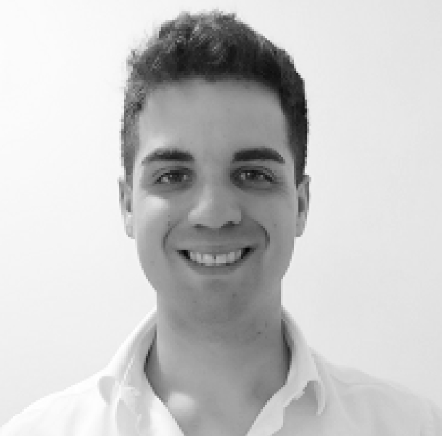
Alessandro Vicentini
Recruiting organisation: Amsterdam University Medical Centers, location University of Amsterdam, Departments of Rehabilitation Medicine and Radiology, Meibergdreef 9, 1105AZ Amsterdam, The Netherlands.
Hosts: Prof. dr. Sicco A. Bus, Prof. dr. Mario Maas
Duration: 42 months
Secondments: Istituto Orthopedico Rizzoli, Bologna, Italy (2 months); Steno Diabetes Center, Copenhagen, Denmark (2 months)
Summary: Foot ulcer recurrence risk in people with diabetes is highest in the early period after healing. This project aims to investigate how the structural and mechanical properties of plantar soft tissues—such as tissue geometry, stiffness, and elasticity—change during this critical remission phase. The doctoral candidate will conduct longitudinal assessments using advanced imaging techniques and plantar pressure analysis to evaluate tissue composition and biomechanics at ulcer healing and during follow-up. By integrating histological, morphological, and mechanical data, the project will provide a deeper understanding of the tissue-level mechanisms underlying ulcer recurrence. These insights are expected to contribute to the development of a comprehensive biomechanical model of plantar tissue recovery, advancing knowledge in diabetic foot disease and informing more effective prevention strategies.

